What is Fibroadenoma Enucleation?
Symptoms of Fibroadenoma
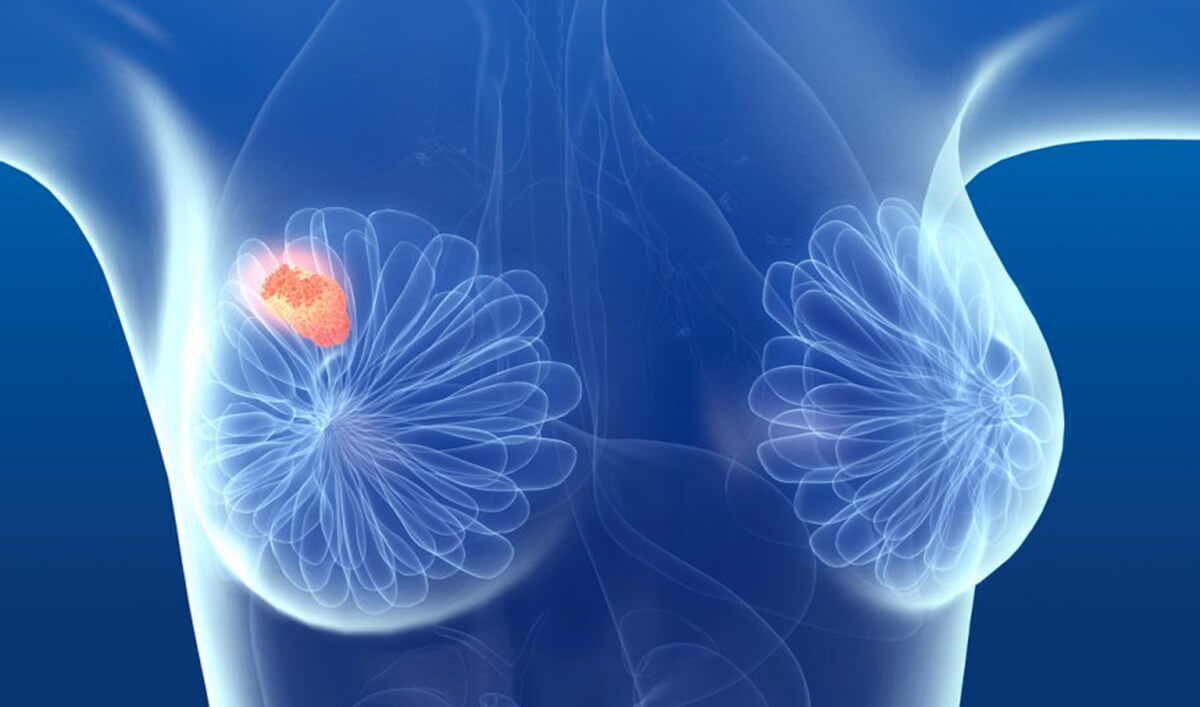
- Presence of a firm, smooth, and rubbery lump in the breast
- Lump that is easily movable under the skin
- Usually painless, but can cause discomfort in some cases
- Size may remain the same or gradually increase
- Typically feels distinct from surrounding breast tissue
Procedure or Treatment for Fibroadenoma Enucleation
Preoperative Assessment:
Breast examination by a surgeon
Ultrasound or mammography to assess the lump
Fine Needle Aspiration Cytology (FNAC) or core needle biopsy to confirm diagnosis
Surgical Procedure:
Performed under local or general anaesthesia
A small incision is made near the lump or along the areola for better cosmetic results
The fibroadenoma is carefully enucleated (shelled out) without removing excess breast tissue
Incision is closed with fine sutures to minimise scarring
Generally done as a day care procedure, allowing same-day discharge
Postoperative Care:
Pain management with analgesics
Minimal restrictions in daily activities
Stitches removal after 7-10 days if non-absorbable sutures are used
Regular follow-up to monitor healing
Prevention
Currently, there are no specific preventive measures for fibroadenomas as their exact cause is not known. However, the following can help in early detection and management:
- Regular breast self-examination
- Routine clinical breast examinations
- Prompt evaluation of any new lump or breast change
Benefits of Fibroadenoma Enucleation
- Removal of lump causing anxiety or discomfort
- Relief from pain or tenderness if present
- Cosmetic improvement of breast contour
- Minimal scarring with careful surgical technique
- Preserves healthy breast tissue and function
- Peace of mind knowing the lump has been removed and confirmed benign by histopathology
Types of Fibroadenomas
Simple Fibroadenoma:
Most common type
Small, painless, and does not increase cancer risk
Complex Fibroadenoma:
Contains other changes like cysts or calcifications
Slightly higher risk of breast cancer than simple fibroadenomas
Giant Fibroadenoma:
Larger than 5 cm or rapidly growing
More common in adolescents and may require removal due to breast distortion
Juvenile Fibroadenoma:
Occurs in girls and adolescents
Often grows rapidly but remains benign